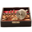
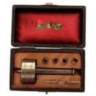
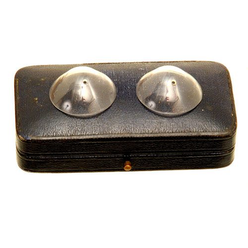
Von Hippel’s automatic corneal trephine, Berlin, c. 1910
On application
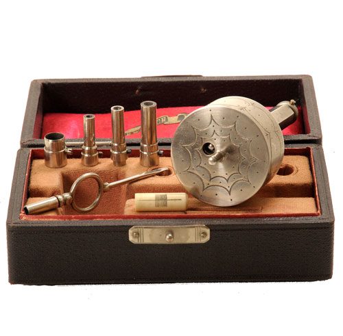
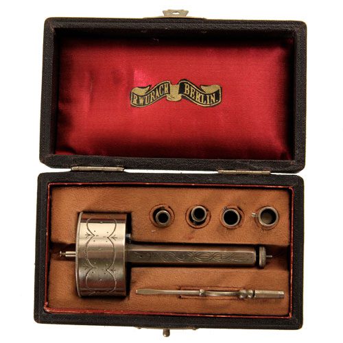
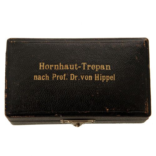
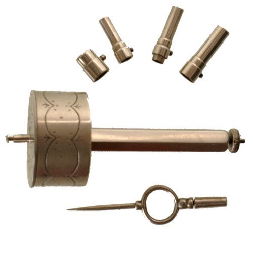
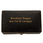
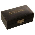
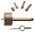
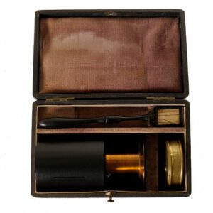
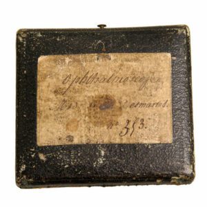
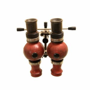
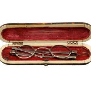
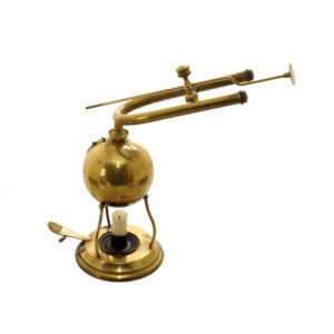
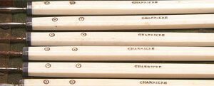
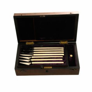
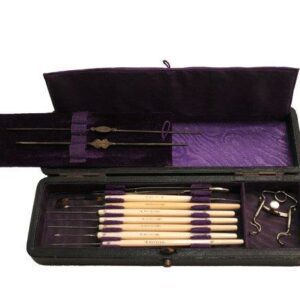
A von Hippel-type automatic corneal trephine, Berlin, c. 1910. The set in original leather box with red velour on the inner side is signed ‘Hornhaut-Trepan nach Prof. Dr. von Hippel’ and Wurach Berlin’. The set is in complete condition, it comes with a wind up drill, two trephine drills, two extensions, a bone cylindrical measuring instrument and a key.
Trephines are normally associated with cutting out a piece of bone from a skull. However, this clockwork trephine was developed to remove a circular piece from the cornea of an eye, to be transplanted into a patient who was experiencing a disease of the cornea, most likely cataracts. Made by R. Wurach Berlin, this trephine was invented by Arthur von Hippel (1841-1916), a German surgeon who experimented using both human and animal subjects to perfect his technique.
It was a significant milestone in keratoplasty: the circular trephine. The device was first popularized by Arthur von Hippel in 1888. It was this trephine that Dr. Eduard Zirm used in his successful corneal transplant on December 7, 1905. Zirm was the chief Opthalmologist of the local hospital at Olmutz, a small Moravian town about 100 miles east of Prague in Slovakia (formerly Czechoslovakia). In this operation Zirm performed two partial penetrating corneal transplants on Alois Glogar, who had been bilaterally blinded by a lime injury 15 months before. Both corneas were opaque except for the extreme periphery, and his vision was limited to hand motions in both eyes.
‘Zirm used the clear cornea of an II-year-old boy, Karl Brauer, who had lost his vision after an intraocular metallic foreign body injury in July 1905. Zirm I had decided to enucleate the sightless and potentially dangerous eye (sympathetic ophthalmia was a serious threat) and to use the clear cornea at the same time for the partial penetrating corneal transplant on Glogar. He was able to harvest two 5-mm corneal buttons using the von Hippel trephine from the same donor cornea.
Under chloroform anesthesia, Glogar had his right cornea trephined with the same 5-mm von Hippel instrument, and a bridge of conjunctiva was sutured over to hold the penetrating graft in place. His left cornea was trephined in a similar manner, but the donor button was taken from a more central part of Brauer’s donated cornea. Zirm fixed this left partial penetrating corneal transplant with two sutures in the conjunctiva, creating a cross over the center of the graft to hold it in place.
The graft on the right eye failed and had to be removed. However, the left corneal transplant cleared. Glogar was released from the hospital 15 weeks after the operation. Antibiotics and steroids were unknown, of course, and diagnostic-related groups were far off. A year later, an ophthalmologist in Fuchs’s clinic checked Glogar’s visual acuity and found it to be 6/36 with a stenopaic disc. He could read 1-4 with a convex glass with difficulty. Zirm died on March 5, 1944, without reporting any other corneal transplants in his 45 publications. Glogar lived for 3 years after the operation.’
This trephine forms the prototype for modern day trephines used in eye surgery, as the same concept of a round trephine is used nowadays, albeit with infinitely improved design and sharpness. Von Hippel is acknowledged as the first surgeon to successfully transplant corneal tissue into a human eye.
In mint condition and dimensions case 10.5 x 6 x 4.5 cm
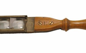
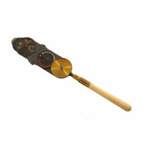
A similar instrument by Weiss is found in the Wellcome collection.
Literature: Peter R. Laibson & Christopher Rapuano, 100- year review of Cornea, in Ophthalmology, 1996
