Jules Cloquet print anatomy of the male human eye 1825
On application
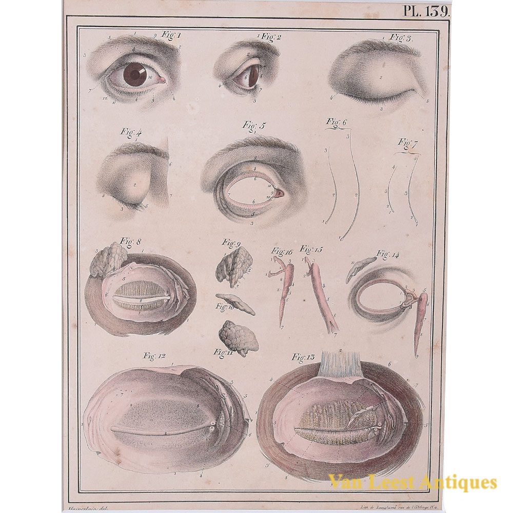
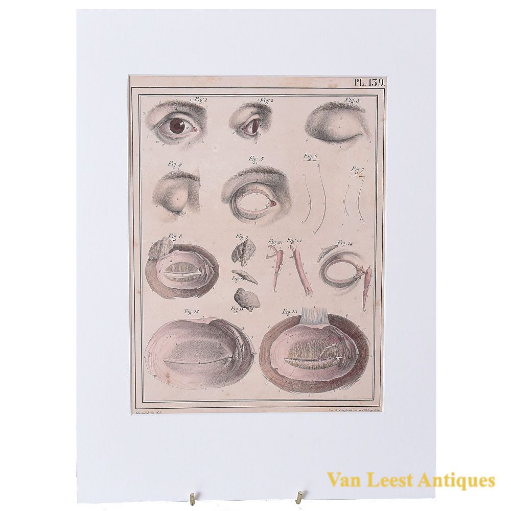
Hand coloured anatomical print no. 139 of the nerves of the eye from Jules Cloquet’s Manuel d’anatomie descriptive du corps humain of 1825 from volume 2 (of 4 volumes). The print shows The right eye of an adult man, seen from the front, the eyelids being open and in profile. The same eye, seen from the front, the eyelids being brought together. Their free edge is slightly turned outwards, in order to show the small holes in which the eyelashes were implanted which are torn out, the orifices of the Meibomian follicles, and the lacrimal points. The Pils torn out of the eyebrows, and increased by four times their size. The eyelashes torn out of the eyelids, and increased in size. Lastly, the eyelids of the left eye with the lacrimal gland, separated from the eye, and seen from behind.
The volume contained over 340 illustrations of Haincelin. Besides from the one shown here, we have many others, with various topics, do not mind to get in touch with us. Jules Germain Cloquet (18 December 1790 – 23 February 1883) was a French physician and surgeon who was born and practiced medicine in Paris. In 1821 Jules Cloquet became one of the earliest members elected to the Académie Nationale de Médecine in Paris. In 1836, he was elected Honorary Fellow of the Royal College of Surgeons in Ireland.
Cloquet was known for his expertise as a surgeon, especially his work with hernial disorders. He was also the first to describe and identify the remnant of the embryonic hyaloid artery. This vestige was to become known as Cloquet’s canal.
Passe-partout dimensions: 37 x 27,5 cm.
