")
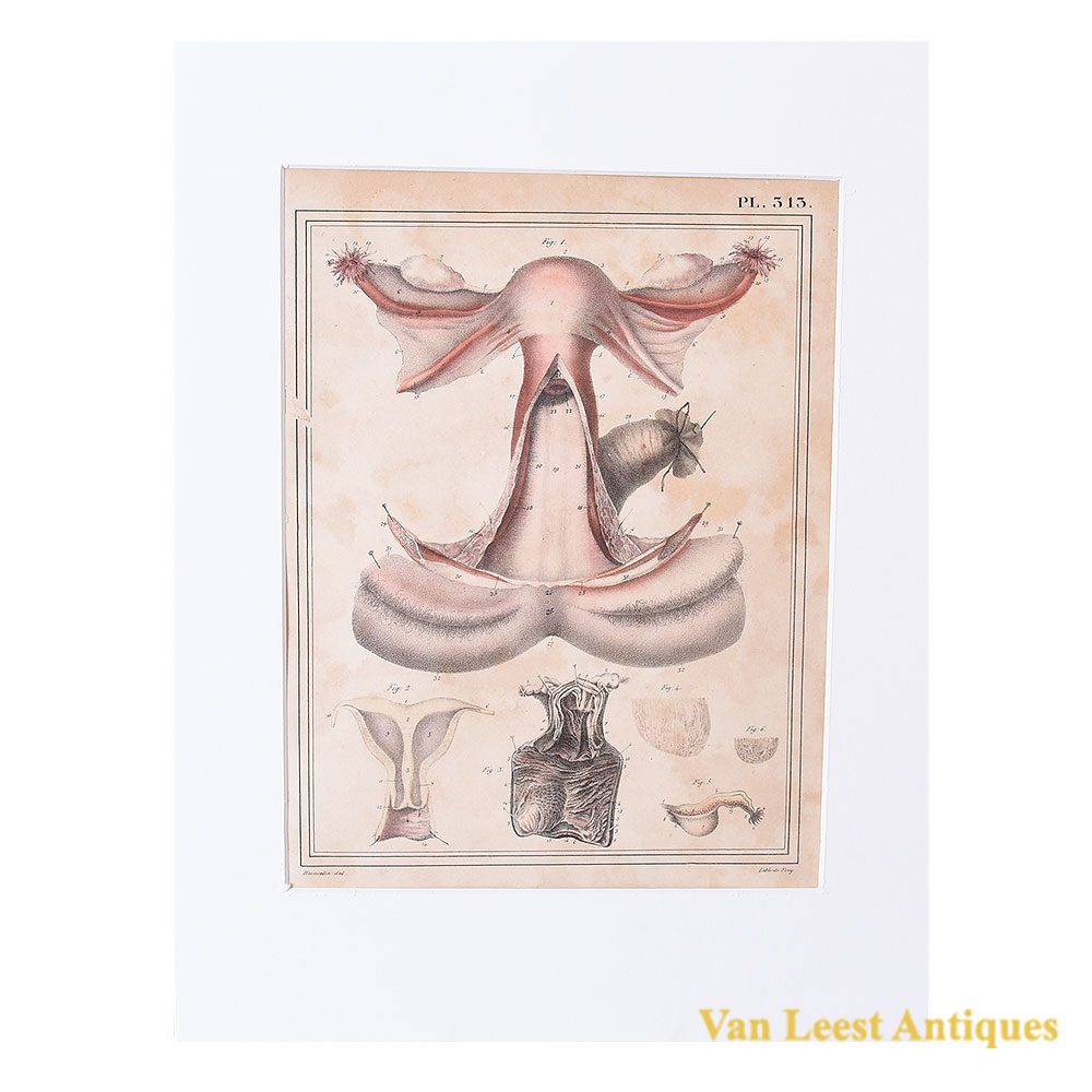

Jules Cloquet print female genital system 1825
Sold
Hand coloured anatomical print no. 313 of the female genital system from Jules Cloquet’s Manuel d’anatomie descriptive du corps humain of 1825 from volume 2 (of 4 volumes). The print representes female genital system; the uterus, ovaries, fallopian tube, round ligaments, and vagina of a twenty-five-year-old woman. These structures are viewed from their anterior side; the ovaries are lifted by retractors and held above their natural position. Only a portion of the peritoneal folds forming the broad ligaments has been preserved. The vagina is longitudinally split along its anterior wall, and the edges of the incision are pulled apart to expose its posterior wall and the cervix, which protrudes into its cavity. The bladder has been removed; the clitoris is cut in the middle, and its two halves are widely separated, as are the corresponding parts of the vulva.
The volume contained over 340 illustrations of Haincelin. Besides from the one shown here, we have many others, with various topics, do not mind to get in touch with us. Jules Germain Cloquet (18 December 1790 – 23 February 1883) was a French physician and surgeon who was born and practiced medicine in Paris. In 1821 Jules Cloquet became one of the earliest members elected to the Académie Nationale de Médecine in Paris. In 1836, he was elected Honorary Fellow of the Royal College of Surgeons in Ireland.
Cloquet was known for his expertise as a surgeon, especially his work with hernial disorders. He was also the first to describe and identify the remnant of the embryonic hyaloid artery. This vestige was to become known as Cloquet’s canal.
Passe-partout dimensions: 37 x 27,5 cm.