")

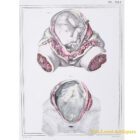

Jules Cloquet print female uterus during pregnancy 1825
€ 155,00
Hand coloured anatomical print no. 321 of the female uterus during pregnancy from Jules Cloquet’s Manuel d’anatomie descriptive du corps humain of 1825 from volume 2 (of 4 volumes). The print representes two states of the uterus, the first represents the fetus in the uterus and in its natural position. The second depicts the uterus in the state of pregnancy, the fetus having been removed. The internal face of the posterior wall of this organ is still covered with membranes: the parts drawn in line have not been numbered, being the same as in the previous figure.
The volume contained over 340 illustrations of Haincelin. Besides from the one shown here, we have many others, with various topics, do not mind to get in touch with us. Jules Germain Cloquet (18 December 1790 – 23 February 1883) was a French physician and surgeon who was born and practiced medicine in Paris. In 1821 Jules Cloquet became one of the earliest members elected to the Académie Nationale de Médecine in Paris. In 1836, he was elected Honorary Fellow of the Royal College of Surgeons in Ireland.
Cloquet was known for his expertise as a surgeon, especially his work with hernial disorders. He was also the first to describe and identify the remnant of the embryonic hyaloid artery. This vestige was to become known as Cloquet’s canal.
Passe-partout dimensions: 37 x 27,5 cm.